Copyright
©2014 Baishideng Publishing Group Inc.
World J Gastroenterol. Jul 28, 2014; 20(28): 9497-9505
Published online Jul 28, 2014. doi: 10.3748/wjg.v20.i28.9497
Published online Jul 28, 2014. doi: 10.3748/wjg.v20.i28.9497
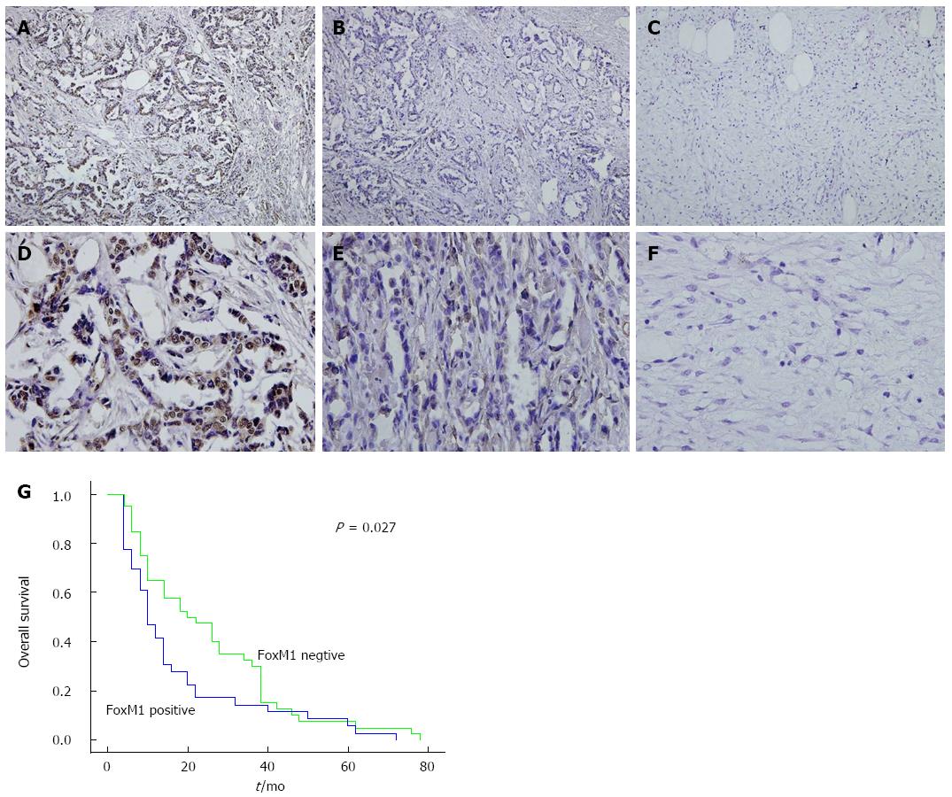
Figure 1 Immunohistochemical analysis of Forkhead box M1 in gallbladder carcinoma (A and D), pericarcinoma (B and E) and healthy tissues (C and F).
Typically, immunohistologic features showed high levels of Forkhead box M1 (FoxM1) expression in carcinoma, low levels of FoxM1 expression in pericarcinoma, and negative staining of FoxM1 in healthy gallbladder tissues. G: Kaplan-Meier analysis of the GBC patients, indicating the poorer survival of the patients with positive FoxM1 expression (P < 0.05). Original magnification (× 100, top; × 400, bottom).
- Citation: Tao J, Xu XS, Song YZ, Qu K, Wu QF, Wang RT, Meng FD, Wei JC, Dong SB, Zhang YL, Tai MH, Dong YF, Wang L, Liu C. Down-regulation of FoxM1 inhibits viability and invasion of gallbladder carcinoma cells, partially dependent on inducement of cellular senescence. World J Gastroenterol 2014; 20(28): 9497-9505
- URL: https://www.wjgnet.com/1007-9327/full/v20/i28/9497.htm
- DOI: https://dx.doi.org/10.3748/wjg.v20.i28.9497


