Copyright
©2014 Baishideng Publishing Group Inc.
World J Gastroenterol. Jul 28, 2014; 20(28): 9476-9485
Published online Jul 28, 2014. doi: 10.3748/wjg.v20.i28.9476
Published online Jul 28, 2014. doi: 10.3748/wjg.v20.i28.9476
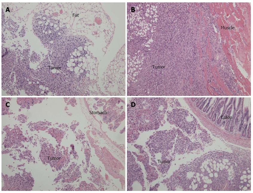
Figure 6 Cross-sections and hematoxylin-eosin staining of intra-abdominal disseminated foci observed under light microscopy (× 100).
A: Tumor implanted in the fat tissue; B: Tumor implanted in the abdominal wall; C: Tumor implanted in the stomach wall; D: Tumor implanted in the colon serosal.
- Citation: Jiang YJ, Lee CL, Wang Q, Zhou ZW, Yang F, Jin C, Fu DL. Establishment of an orthotopic pancreatic cancer mouse model: Cells suspended and injected in Matrigel. World J Gastroenterol 2014; 20(28): 9476-9485
- URL: https://www.wjgnet.com/1007-9327/full/v20/i28/9476.htm
- DOI: https://dx.doi.org/10.3748/wjg.v20.i28.9476


