Copyright
©2014 Baishideng Publishing Group Inc.
World J Gastroenterol. Jul 14, 2014; 20(26): 8729-8735
Published online Jul 14, 2014. doi: 10.3748/wjg.v20.i26.8729
Published online Jul 14, 2014. doi: 10.3748/wjg.v20.i26.8729
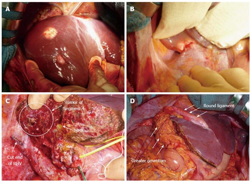
Figure 2 Intraoperative images.
A: The tumors were well-circumscribed, whitish, and firm; B: The tumor of segment 6 invaded Gerota’s fascia; C: After right lobectomy, the tumor of segment 4 was very close to the cut surface of the liver; D: A flap of the greater omentum (arrows) was sutured over the cut surface of the liver to cover the tumor of segment 4. MHV: Middle hepatic vein; RHV: Right hepatic vein.
- Citation: Komatsu S, Iwasaki T, Demizu Y, Terashima K, Fujii O, Takebe A, Toyokawa A, Teramura K, Fukumoto T, Ku Y, Fuwa N. Two-stage treatment with hepatectomy and carbon-ion radiotherapy for multiple hepatic epithelioid hemangioendotheliomas. World J Gastroenterol 2014; 20(26): 8729-8735
- URL: https://www.wjgnet.com/1007-9327/full/v20/i26/8729.htm
- DOI: https://dx.doi.org/10.3748/wjg.v20.i26.8729


