Copyright
©2014 Baishideng Publishing Group Inc.
World J Gastroenterol. Jul 14, 2014; 20(26): 8729-8735
Published online Jul 14, 2014. doi: 10.3748/wjg.v20.i26.8729
Published online Jul 14, 2014. doi: 10.3748/wjg.v20.i26.8729
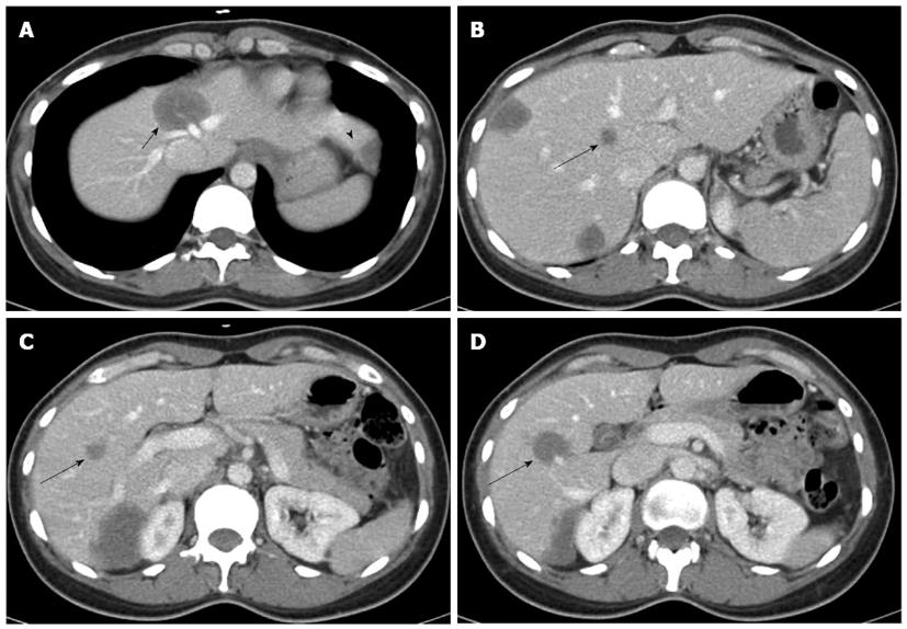
Figure 1 Abdominal computed tomography revealed a total of 7 lesions located in both lobes.
A: The tumor of segment 4 (short arrow) hinders the possibility of right hemihepatectomy, because it invades the roots of the middle and left hepatic veins (arrowhead indicates the tumor of segment 3); B-D: The 3 deep-seated tumors in the right lobe (long arrows) hinder the possibility of left hemihepatectomy.
- Citation: Komatsu S, Iwasaki T, Demizu Y, Terashima K, Fujii O, Takebe A, Toyokawa A, Teramura K, Fukumoto T, Ku Y, Fuwa N. Two-stage treatment with hepatectomy and carbon-ion radiotherapy for multiple hepatic epithelioid hemangioendotheliomas. World J Gastroenterol 2014; 20(26): 8729-8735
- URL: https://www.wjgnet.com/1007-9327/full/v20/i26/8729.htm
- DOI: https://dx.doi.org/10.3748/wjg.v20.i26.8729


