Copyright
©2014 Baishideng Publishing Group Inc.
World J Gastroenterol. Jul 14, 2014; 20(26): 8449-8457
Published online Jul 14, 2014. doi: 10.3748/wjg.v20.i26.8449
Published online Jul 14, 2014. doi: 10.3748/wjg.v20.i26.8449
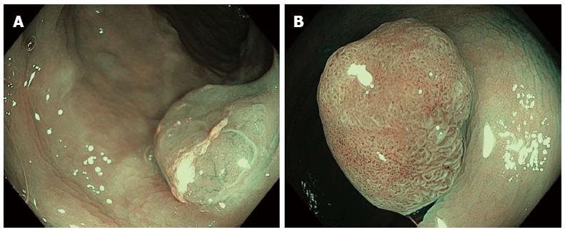
Figure 5 High-resolution endoscopic image of narrow band imaging.
A: Hyperplastic polyp with weak vascular pattern intensity and type II Kudo’s pit pattern classification; B: Tubular adenoma with foci of high grade dysplasia exhibiting strong vascular pattern intensity and type IIIL Kudo’s pit pattern classification.
- Citation: Lopez-Ceron M, Sanabria E, Pellise M. Colonic polyps: Is it useful to characterize them with advanced endoscopy? World J Gastroenterol 2014; 20(26): 8449-8457
- URL: https://www.wjgnet.com/1007-9327/full/v20/i26/8449.htm
- DOI: https://dx.doi.org/10.3748/wjg.v20.i26.8449


