Copyright
©2014 Baishideng Publishing Group Inc.
World J Gastroenterol. Jul 7, 2014; 20(25): 8317-8319
Published online Jul 7, 2014. doi: 10.3748/wjg.v20.i25.8317
Published online Jul 7, 2014. doi: 10.3748/wjg.v20.i25.8317
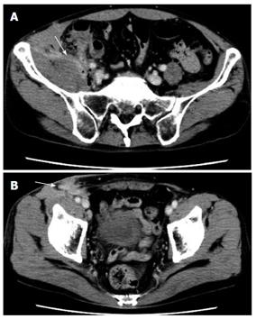
Figure 1 Abdominal contrast computed tomography.
A: The psoas abscess (white arrow) was adjacent to the swollen appendix; B: A cutaneous fistula (white arrow) was observed in the right inguinal region.
- Citation: Otowa Y, Sumi Y, Kanaji S, Kanemitsu K, Yamashita K, Imanishi T, Nakamura T, Suzuki S, Tanaka K, Kakeji Y. Appendicitis with psoas abscess successfully treated by laparoscopic surgery. World J Gastroenterol 2014; 20(25): 8317-8319
- URL: https://www.wjgnet.com/1007-9327/full/v20/i25/8317.htm
- DOI: https://dx.doi.org/10.3748/wjg.v20.i25.8317


