Copyright
©2014 Baishideng Publishing Group Inc.
World J Gastroenterol. Jul 7, 2014; 20(25): 8304-8308
Published online Jul 7, 2014. doi: 10.3748/wjg.v20.i25.8304
Published online Jul 7, 2014. doi: 10.3748/wjg.v20.i25.8304
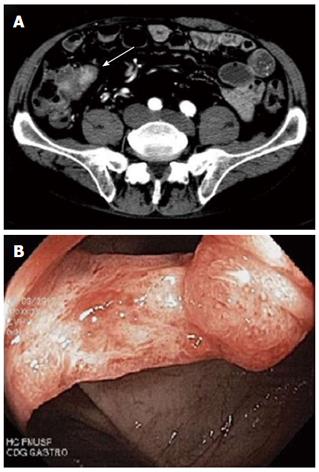
Figure 1 Imaging features.
A: Abdominal computed tomography showing a thickening of the terminal ileum (white arrow); B: Oedematous and friable ileocaecal valve with an infiltrative lesion observed during colonoscopy.
- Citation: Lugao RDS, Motta MP, Azevedo MFC, Lima RGR, Abrantes FA, Abdala E, Carrilho FJ, Mazo DFC. Immune thrombocytopenic purpura induced by intestinal tuberculosis in a liver transplant recipient. World J Gastroenterol 2014; 20(25): 8304-8308
- URL: https://www.wjgnet.com/1007-9327/full/v20/i25/8304.htm
- DOI: https://dx.doi.org/10.3748/wjg.v20.i25.8304


