Copyright
©2014 Baishideng Publishing Group Inc.
World J Gastroenterol. Jun 28, 2014; 20(24): 7864-7877
Published online Jun 28, 2014. doi: 10.3748/wjg.v20.i24.7864
Published online Jun 28, 2014. doi: 10.3748/wjg.v20.i24.7864
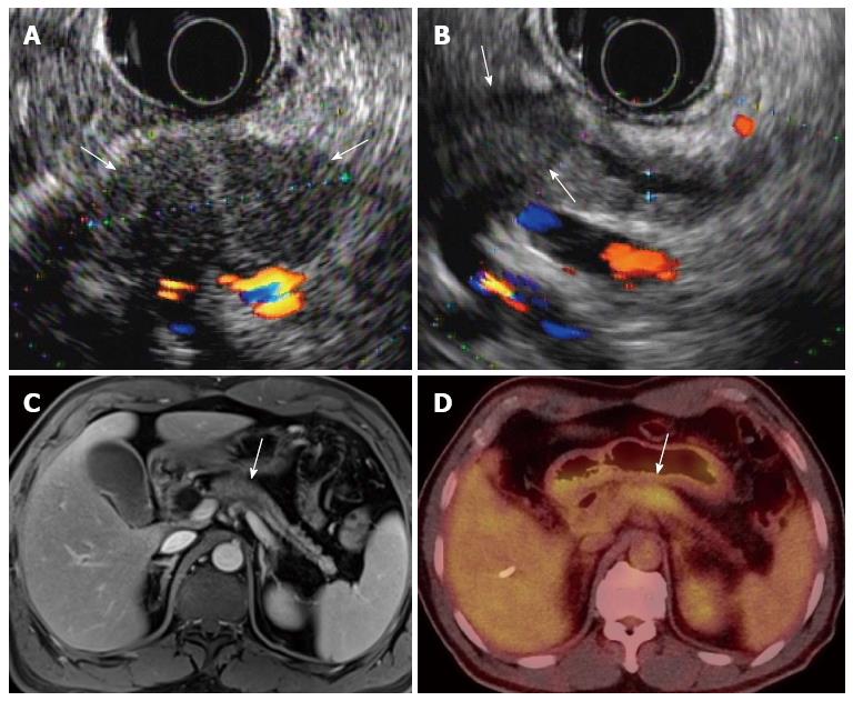
Figure 1 A 58-year-old, male patient with pancreatic body cancer with typical imaging findings.
A, B: Endoscopic ultrasonography shows an approximately 3-cm, hypoechoic mass (arrows) in the pancreatic body with distal pancreatic duct dilatation; C, D: The mass (arrow) shows hypointensity on portal-venous-phase, contrast enhanced MR and hypermetabolism on PET/CT, respectively. MR: Magnetic resonance; PET: Positron emission tomography; CT: Computed tomography.
- Citation: Lee ES, Lee JM. Imaging diagnosis of pancreatic cancer: A state-of-the-art review. World J Gastroenterol 2014; 20(24): 7864-7877
- URL: https://www.wjgnet.com/1007-9327/full/v20/i24/7864.htm
- DOI: https://dx.doi.org/10.3748/wjg.v20.i24.7864


