Copyright
©2014 Baishideng Publishing Group Inc.
World J Gastroenterol. Jun 28, 2014; 20(24): 7785-7793
Published online Jun 28, 2014. doi: 10.3748/wjg.v20.i24.7785
Published online Jun 28, 2014. doi: 10.3748/wjg.v20.i24.7785
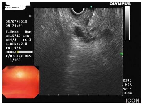
Figure 4 Fine-needle aspiration of a branch-duct intraductal papillary mucinous neoplasms.
Material obtained was mucinous (Papanikolaou staining) with low cellularity. Mucinous cells did not demonstrate nuclear atypia and expressed MUC5AC, but not MUC1 or MUC2 on immunostaining, findings consistent with a benign branch-duct intraductal papillary mucinous neoplasms.
- Citation: Efthymiou A, Podas T, Zacharakis E. Endoscopic ultrasound in the diagnosis of pancreatic intraductal papillary mucinous neoplasms. World J Gastroenterol 2014; 20(24): 7785-7793
- URL: https://www.wjgnet.com/1007-9327/full/v20/i24/7785.htm
- DOI: https://dx.doi.org/10.3748/wjg.v20.i24.7785


