Copyright
©2014 Baishideng Publishing Group Inc.
World J Gastroenterol. Jun 21, 2014; 20(23): 7452-7460
Published online Jun 21, 2014. doi: 10.3748/wjg.v20.i23.7452
Published online Jun 21, 2014. doi: 10.3748/wjg.v20.i23.7452
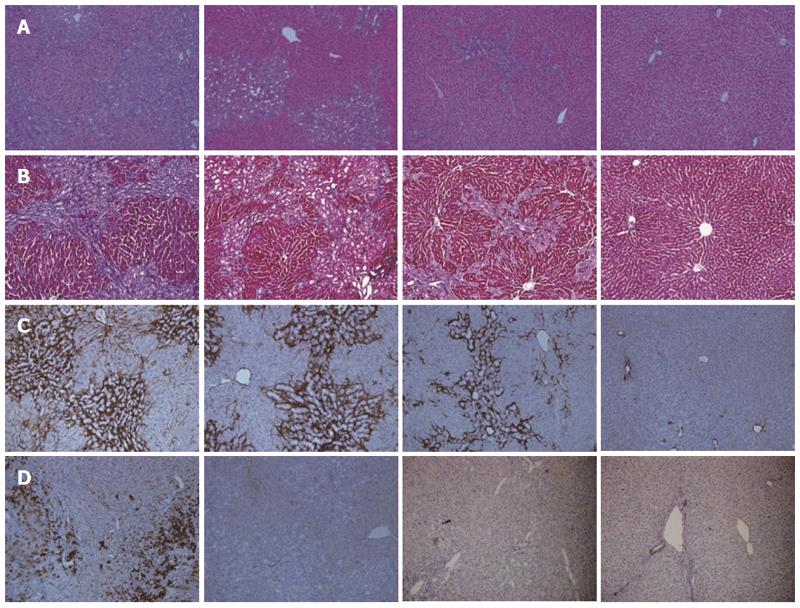
Figure 1 Comparison of histological findings between the groups that underwent bile duct ligation (BDL) with/without rapamycin treatment and the control.
A: Hematoxylin and eosin (HE) staining. The bile duct ligation (BDL)+/Rapa++ group showed the smallest expansion of portal areas, with the least amount of ductular proliferation and the most mild inflammatory reaction among the BDL groups; B: Masson-Trichrome staining. The BDL+/Rapa++ group showed the lowest degree of fibrotic septa formation and deposition of collagen among BDL groups (blue); C: α-smooth muscle actin (α-SMA) staining (dark brown). The BDL+/Rapa++ group showed the lowest amount of staining among BDL groups; D: Cytokeratin 19 (CK 19) protein expression (dark brown). The BDL+/Rapa++ group showed the lowest amount of staining among BDL groups. Rapa: Rapamycin; Rapa+: Rat administered rapamycin for 28 d from the 14th day post-BDL; Rapa++: Rat administered rapamycin for 28 d beginning the first day post-BDL. Original magnification: × 100(a-d).
- Citation: Kim YJ, Lee ES, Kim SH, Lee HY, Noh SM, Kang DY, Lee BS. Inhibitory effects of rapamycin on the different stages of hepatic fibrosis. World J Gastroenterol 2014; 20(23): 7452-7460
- URL: https://www.wjgnet.com/1007-9327/full/v20/i23/7452.htm
- DOI: https://dx.doi.org/10.3748/wjg.v20.i23.7452


