Copyright
©2014 Baishideng Publishing Group Inc.
World J Gastroenterol. Jun 21, 2014; 20(23): 7442-7451
Published online Jun 21, 2014. doi: 10.3748/wjg.v20.i23.7442
Published online Jun 21, 2014. doi: 10.3748/wjg.v20.i23.7442
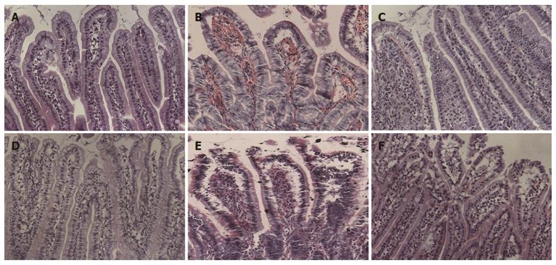
Figure 2 Graft histopathology at different time points after heterotopic intestinal transplantation (HE staining, × 200).
A: Normal intestine with normal villous architecture and glands in group A; B: Intestinal mucosa degradation and hemorrhage of the lamina propria, ulceration, decreased ratio of villus height and crypt height, aggravated lymphocyte infiltration, partial gland epithelial necrosis in group B at day 5 after operation; D: There were decreased intestinal mucosal villi with mild deformity and interstitial infiltration of inflammatory cells; E: The condition was aggravated 7 d after heterotopic intestinal transplantation, and there was epithelial degeneration, intestinal wall thinning and necrosis, and interstitial inflammatory cell infiltration in great quantities; C, F: Recovery of the damaged mucosa in group A at (C) day 5 and (F) day 7.
- Citation: Zhang W, Shen ZY, Song HL, Yang Y, Wu BJ, Fu NN, Liu T. Protective effect of bone marrow mesenchymal stem cells in intestinal barrier permeability after heterotopic intestinal transplantation. World J Gastroenterol 2014; 20(23): 7442-7451
- URL: https://www.wjgnet.com/1007-9327/full/v20/i23/7442.htm
- DOI: https://dx.doi.org/10.3748/wjg.v20.i23.7442


