Copyright
©2014 Baishideng Publishing Group Inc.
World J Gastroenterol. Jun 14, 2014; 20(22): 7075-7078
Published online Jun 14, 2014. doi: 10.3748/wjg.v20.i22.7075
Published online Jun 14, 2014. doi: 10.3748/wjg.v20.i22.7075
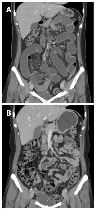
Figure 2 Follow up computed tomography image after the patient developed mechanical ileus.
A: One month before the operation. Follow up computed tomography (CT) demonstrated a transitional zone at the distal ileum near the pelvic abscess, with small bowel dilatation; B: Seven day before the operation. Follow-up CT showed a linear, radiopaque lesion that was partially penetrated into the dome of the bladder.
- Citation: Cho MK, Lee MS, Han HY, Woo SH. Fish bone migration to the urinary bladder after rectosigmoid colon perforation. World J Gastroenterol 2014; 20(22): 7075-7078
- URL: https://www.wjgnet.com/1007-9327/full/v20/i22/7075.htm
- DOI: https://dx.doi.org/10.3748/wjg.v20.i22.7075


