Copyright
©2014 Baishideng Publishing Group Inc.
World J Gastroenterol. Jun 14, 2014; 20(22): 7061-7066
Published online Jun 14, 2014. doi: 10.3748/wjg.v20.i22.7061
Published online Jun 14, 2014. doi: 10.3748/wjg.v20.i22.7061
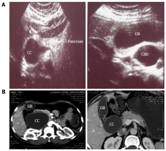
Figure 1 Ultrasound imaging and abdominal computed tomography of type I choledochal cyst.
A: Ultrasound imaging; B: Abdominal CT. CC: Choledochal cyst; GB: Gall bladder; CBD: Common bile duct; CT: Computed tomography.
- Citation: Gadelhak N, Shehta A, Hamed H. Diagnosis and management of choledochal cyst: 20 years of single center experience. World J Gastroenterol 2014; 20(22): 7061-7066
- URL: https://www.wjgnet.com/1007-9327/full/v20/i22/7061.htm
- DOI: https://dx.doi.org/10.3748/wjg.v20.i22.7061


