Copyright
©2014 Baishideng Publishing Group Inc.
World J Gastroenterol. Jun 7, 2014; 20(21): 6685-6690
Published online Jun 7, 2014. doi: 10.3748/wjg.v20.i21.6685
Published online Jun 7, 2014. doi: 10.3748/wjg.v20.i21.6685
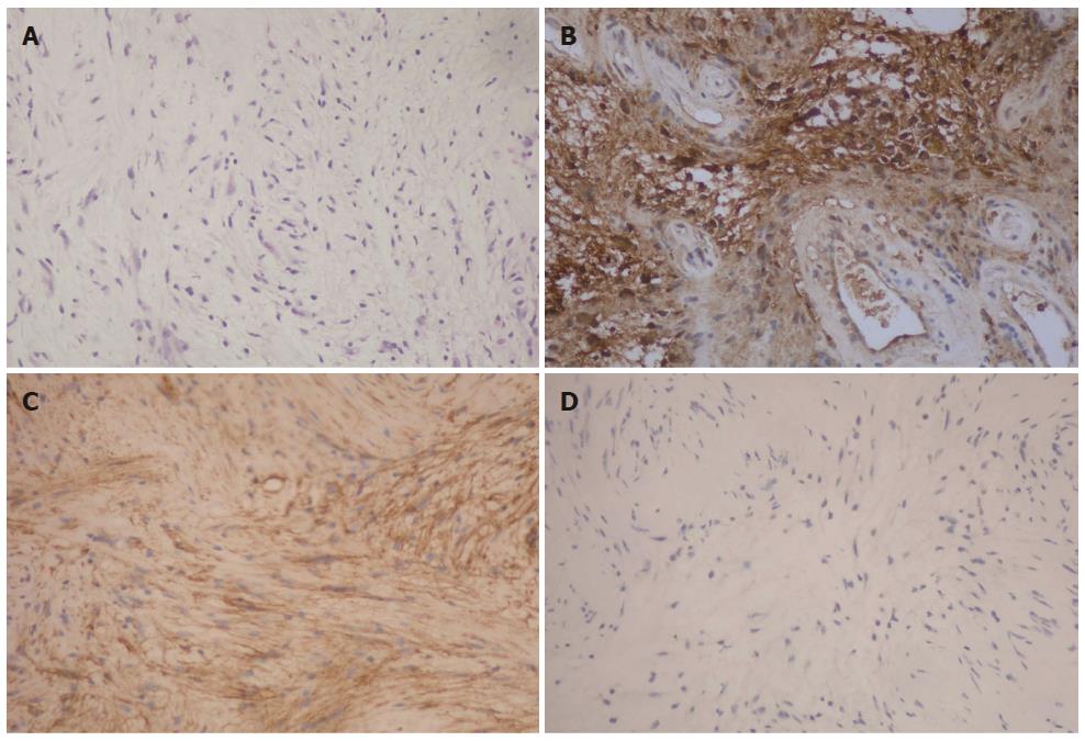
Figure 3 Pathological examinations.
A: Microscopic examination shows that the tumor mainly composes of spindle-shaped cells and no atypical cells are found (HE staining; magnification: 10 × 30); B, C, D: Immunohistochemical staining shows the tumor is positive for S-100 protein (10 × 30) (B) and CD56(10 × 30) (C), whereas negative for CD117 (10 × 30) (D).
- Citation: Liu LN, Xu HX, Zheng SG, Sun LP, Guo LH, Wu J. Solitary schwannoma of the gallbladder: A case report and literature review. World J Gastroenterol 2014; 20(21): 6685-6690
- URL: https://www.wjgnet.com/1007-9327/full/v20/i21/6685.htm
- DOI: https://dx.doi.org/10.3748/wjg.v20.i21.6685


