Copyright
©2014 Baishideng Publishing Group Inc.
World J Gastroenterol. Jun 7, 2014; 20(21): 6651-6657
Published online Jun 7, 2014. doi: 10.3748/wjg.v20.i21.6651
Published online Jun 7, 2014. doi: 10.3748/wjg.v20.i21.6651
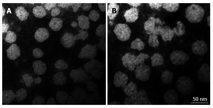
Figure 1 Characterization of the exosomes of Huh-7 cells extracted by two methods.
Electron micrographs of exosomes isolated from Huh-7 cells in serum-free concentrated culture medium by the traditional method (A) and the new extraction method based on a nanomaterial (B). Negative-stained images show exosomes with a smooth, saucer-like morphology. The sizes are between 30-100 nm. The scale bar is 50 nm. Direct Mag: 100000 ×, HV = 80.0 kV.
- Citation: Zhu L, Qu XH, Sun YL, Qian YM, Zhao XH. Novel method for extracting exosomes of hepatocellular carcinoma cells. World J Gastroenterol 2014; 20(21): 6651-6657
- URL: https://www.wjgnet.com/1007-9327/full/v20/i21/6651.htm
- DOI: https://dx.doi.org/10.3748/wjg.v20.i21.6651


