Copyright
©2014 Baishideng Publishing Group Inc.
World J Gastroenterol. Jun 7, 2014; 20(21): 6523-6533
Published online Jun 7, 2014. doi: 10.3748/wjg.v20.i21.6523
Published online Jun 7, 2014. doi: 10.3748/wjg.v20.i21.6523
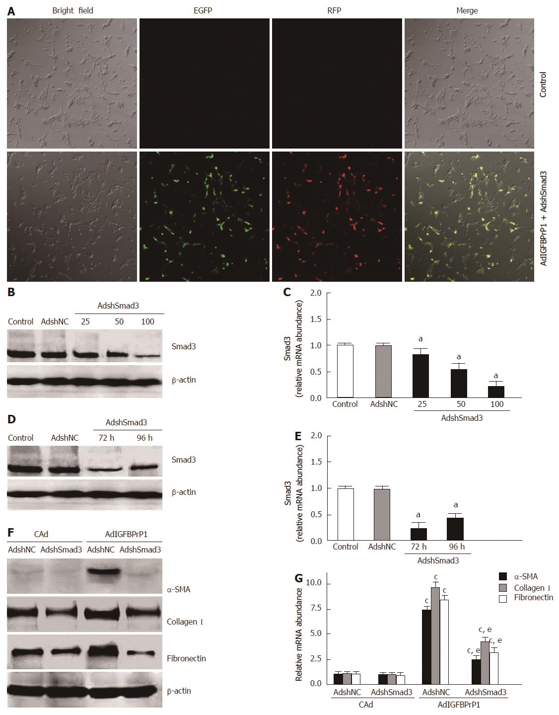
Figure 3 Insulin-like growth factor binding protein-related protein 1-induced extracellular matrix expression is mediated through the Smad pathway in hepatic stellate cell-T6 cells.
Hepatic stellate cell-T6 (HSC-T6) cells were co-infected with adenovirus vectors containing shSmad3 (AdshSmad3) or shNC (AdshNC) and adenovirus vector carrying insulin-like growth factor binding protein-related protein 1 (IGFBPrP1) (AdIGFBPrP1). A: Expression of enhanced green fluorescent protein (EGFP) and red fluorescent protein (RFP) in HSC-T6 cells was visualized by confocal microscopy after treatment with AdshSmad3 (magnification ×200); B, C: Smad3 protein (B) and mRNA (C) expression in HSC-T6 cells was detected by Western blot and real-time polymerase chain reaction (RT-PCR) after treatment with different multiplicity of infection (MOI) of AdshSmad3, respectively; D, E: Smad3 protein (D) and mRNA (E) expression was detected by Western blot and real-time RT-PCR 72 h or 96 h after AdshSmad3 treatment (MOI = 100), respectively; F, G: Protein (F) and mRNA (G) expression of α-smooth muscle actin (α-SMA) and extracellular matrix in HSC-T6 cells was analyzed by Western blot and real-time RT-PCR 72 h after AdshSmad3 treatment (MOI = 100), respectively. Data are expressed as mean ± SD (n = 4 per group). aP < 0.05 vs the levels in the control group; cP < 0.05 vs the levels in cAd + AdshNC; eP < 0.05 vs the levels in AdIGFBPrP1 + AdshNC.
-
Citation: Zhang Y, Zhang QQ, Guo XH, Zhang HY, Liu LX. IGFBPrP1 induces liver fibrosis by inducing hepatic stellate cell activation and hepatocyte apoptosis
via Smad2/3 signaling. World J Gastroenterol 2014; 20(21): 6523-6533 - URL: https://www.wjgnet.com/1007-9327/full/v20/i21/6523.htm
- DOI: https://dx.doi.org/10.3748/wjg.v20.i21.6523


