Copyright
©2014 Baishideng Publishing Group Inc.
World J Gastroenterol. Jun 7, 2014; 20(21): 6420-6424
Published online Jun 7, 2014. doi: 10.3748/wjg.v20.i21.6420
Published online Jun 7, 2014. doi: 10.3748/wjg.v20.i21.6420
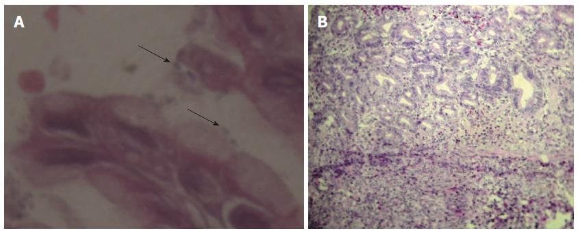
Figure 2 Antral mucosa from Helicobacter pylori-infected rats with hematoxylin and eosin staining.
A: Helicobacter pylori organism in the gastric mucosa (arrow) (600 ×); B: Gastric mucosa with erosion and scattered infiltration of inflammatory cells (250 ×).
-
Citation: Werawatganon D. Simple animal model of
Helicobacter pylori infection. World J Gastroenterol 2014; 20(21): 6420-6424 - URL: https://www.wjgnet.com/1007-9327/full/v20/i21/6420.htm
- DOI: https://dx.doi.org/10.3748/wjg.v20.i21.6420


