Copyright
©2014 Baishideng Publishing Group Inc.
World J Gastroenterol. May 28, 2014; 20(20): 6180-6200
Published online May 28, 2014. doi: 10.3748/wjg.v20.i20.6180
Published online May 28, 2014. doi: 10.3748/wjg.v20.i20.6180
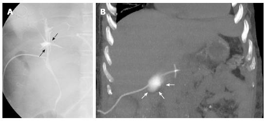
Figure 2 T-tube cholangiography.
A: The examination performed few days after liver transplant in a 28-year-old female with primary sclerosing cholangitis shows bile leakage at the anastomotic site (black arrows); B: Fifty-six year-old male recipient. Coronal maximum intensity projection reconstruction from unenhanced multidetector computed tomography scan shows contrast medium extravasation in the right subhepatic space, suggesting T-tube rupture (white arrows).
- Citation: Girometti R, Como G, Bazzocchi M, Zuiani C. Post-operative imaging in liver transplantation: State-of-the-art and future perspectives. World J Gastroenterol 2014; 20(20): 6180-6200
- URL: https://www.wjgnet.com/1007-9327/full/v20/i20/6180.htm
- DOI: https://dx.doi.org/10.3748/wjg.v20.i20.6180


