Copyright
©2014 Baishideng Publishing Group Co.
World J Gastroenterol. Jan 14, 2014; 20(2): 603-606
Published online Jan 14, 2014. doi: 10.3748/wjg.v20.i2.603
Published online Jan 14, 2014. doi: 10.3748/wjg.v20.i2.603
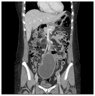
Figure 1 A contrast-enhanced coronal image of computed tomography reveals a well-circumscribed and poorly attenuated dumbbell-shaped mass exhibiting peripheral enhancement and a trilaminar appearance in the pelvic cavity.
- Citation: Park JY, Her KH, Kim BS, Maeng YH. A completely isolated intestinal duplication cyst mimicking ovarian cyst torsion in an adult. World J Gastroenterol 2014; 20(2): 603-606
- URL: https://www.wjgnet.com/1007-9327/full/v20/i2/603.htm
- DOI: https://dx.doi.org/10.3748/wjg.v20.i2.603


