Copyright
©2014 Baishideng Publishing Group Inc.
World J Gastroenterol. May 21, 2014; 20(19): 5918-5923
Published online May 21, 2014. doi: 10.3748/wjg.v20.i19.5918
Published online May 21, 2014. doi: 10.3748/wjg.v20.i19.5918
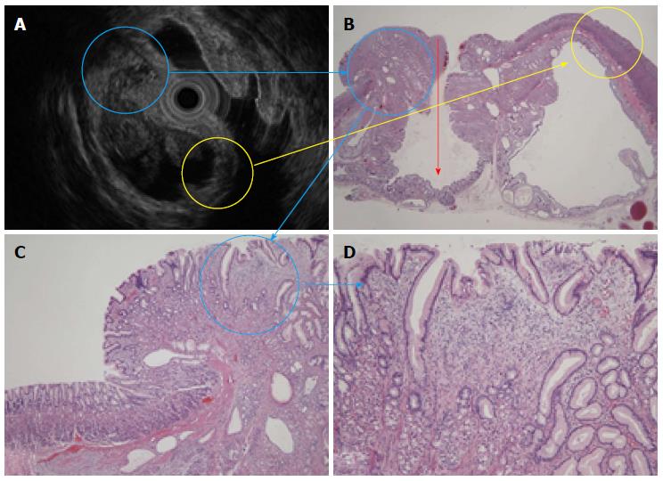
Figure 5 An endoscopic ultrasound and histological comparison.
A: An endoscopic ultrasound (EUS) revealed a heterogeneous tumor; B: The 5 layers shown in the EUS and HE stain (× 20) (yellow circle) consisted of a normal mucosa layer, immature fibroblast cells, pyloric glands, muscularis mucosa, and another normal mucosa layer. The surface mucosa was inverted into the submucosal layer (red arrow); C and D: The hyperechoic solid portion had tiny low echoic cystic spots (blue circle) and showed the proliferation of pseudo-pylorus glands, cystic glands, fibroblast cells, smooth muscle, and nerve elements.
- Citation: Mori H, Kobara H, Tsushimi T, Fujihara S, Nishiyama N, Matsunaga T, Ayaki M, Yachida T, Masaki T. Two rare gastric hamartomatous inverted polyp cases suggest the pathogenesis of growth. World J Gastroenterol 2014; 20(19): 5918-5923
- URL: https://www.wjgnet.com/1007-9327/full/v20/i19/5918.htm
- DOI: https://dx.doi.org/10.3748/wjg.v20.i19.5918


