Copyright
©2014 Baishideng Publishing Group Inc.
World J Gastroenterol. May 21, 2014; 20(19): 5918-5923
Published online May 21, 2014. doi: 10.3748/wjg.v20.i19.5918
Published online May 21, 2014. doi: 10.3748/wjg.v20.i19.5918
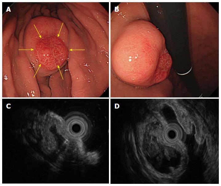
Figure 3 An submucosal tumor measuring 40 mm in diameter was found in the greater curvature of the middle body.
A and B: The size of the submucosal tumor (SMT) had increased from the 20 mm in diameter that was reported 5 years before. In addition, a reddish erosive part 10 mm in diameter was found on the top of the SMT (yellow arrows). C and D: An endoscopic ultrasound revealed a heterogeneous tumor with small spotty cystic areas, a large anechoic cystic part, with papillary structures and 5 specific structural layers as its wall.
- Citation: Mori H, Kobara H, Tsushimi T, Fujihara S, Nishiyama N, Matsunaga T, Ayaki M, Yachida T, Masaki T. Two rare gastric hamartomatous inverted polyp cases suggest the pathogenesis of growth. World J Gastroenterol 2014; 20(19): 5918-5923
- URL: https://www.wjgnet.com/1007-9327/full/v20/i19/5918.htm
- DOI: https://dx.doi.org/10.3748/wjg.v20.i19.5918


