Copyright
©2014 Baishideng Publishing Group Inc.
World J Gastroenterol. May 21, 2014; 20(19): 5918-5923
Published online May 21, 2014. doi: 10.3748/wjg.v20.i19.5918
Published online May 21, 2014. doi: 10.3748/wjg.v20.i19.5918
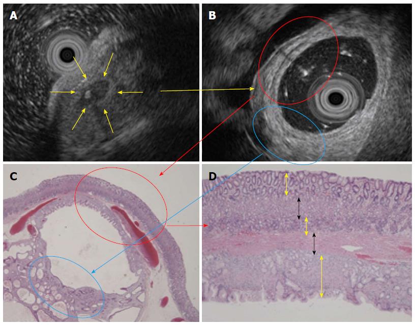
Figure 2 Endoscopic ultrasound before and after the biopsy and histology.
A: An EUS before biopsy revealed a heterogeneous tumor with a cystic area and calcification spot (yellow arrow); B: An endoscopic ultrasound (EUS) from inside the submucosal tumor (SMT) filled with distilled water and an inserted EUS probe showed 5 specific layers (red circle); C: An HE stain (× 20) also showed the 5 structural layers. The hyperechoic portion showed proliferation of pseudo-pylorus glands and cystic gland dilatation without cytological atypia (blue circle); D: The 5 layers consisted of a normal mucosal layer (yellow arrow), immature fibroblasts (black arrow), pyloric glands (yellow arrow), muscularis mucosa (black arrow) and another normal mucosal layer (yellow arrow) (HE stain × 100).
- Citation: Mori H, Kobara H, Tsushimi T, Fujihara S, Nishiyama N, Matsunaga T, Ayaki M, Yachida T, Masaki T. Two rare gastric hamartomatous inverted polyp cases suggest the pathogenesis of growth. World J Gastroenterol 2014; 20(19): 5918-5923
- URL: https://www.wjgnet.com/1007-9327/full/v20/i19/5918.htm
- DOI: https://dx.doi.org/10.3748/wjg.v20.i19.5918


