Copyright
©2014 Baishideng Publishing Group Inc.
World J Gastroenterol. May 21, 2014; 20(19): 5859-5866
Published online May 21, 2014. doi: 10.3748/wjg.v20.i19.5859
Published online May 21, 2014. doi: 10.3748/wjg.v20.i19.5859
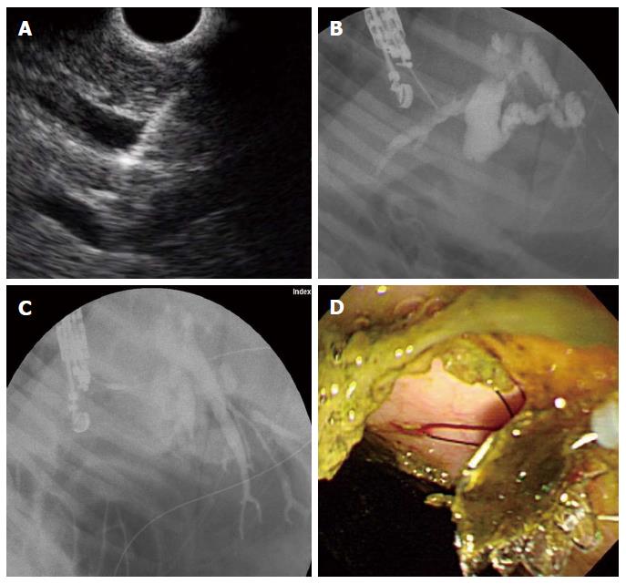
Figure 4 Endoscopic ultrasound-guided hepaticoenterostomy.
A: Linear endoscopic ultrasound (EUS) image shows a 19-G needle puncture targeting the intrahepatic bile duct (IHD; B: Fluoroscopic image reveals marked IHD and common bile duct dilatation with ampullary obstruction; C: After EUS-guided 19-G needle puncture of the IHD, the device for one-step endoscopic ultrasound-guided biliary drainage system was inserted and the stent released under the guidance of EUS and fluoroscopy; D: Following successful deployment of the stent, the bile passage was seen in the high body of stomach.
- Citation: Lee TH, Choi JH, Lee SS, Cho HD, Seo DW, Park SH, Lee SK, Kim MH, Park DH. A pilot proof-of-concept study of a modified device for one-step endoscopic ultrasound-guided biliary drainage in a new experimental biliary dilatation animal model. World J Gastroenterol 2014; 20(19): 5859-5866
- URL: https://www.wjgnet.com/1007-9327/full/v20/i19/5859.htm
- DOI: https://dx.doi.org/10.3748/wjg.v20.i19.5859


