Copyright
©2014 Baishideng Publishing Group Inc.
World J Gastroenterol. May 21, 2014; 20(19): 5849-5858
Published online May 21, 2014. doi: 10.3748/wjg.v20.i19.5849
Published online May 21, 2014. doi: 10.3748/wjg.v20.i19.5849
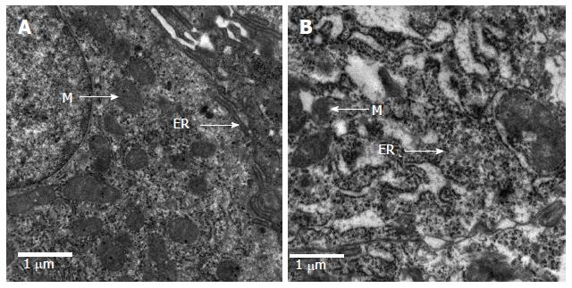
Figure 4 Ultra-structural analysis of intestinal mucosa in rats.
Tissue samples were fixed, cut, stained and examined by transmission electron microscopy. Images are representative electron micrographs of the normal (A) and the model (B) groups. Cell organelles: ER: Endoplasmic reticulum; M: Mitochondria.
- Citation: Li Y, Liu XY, Ma MM, Qi ZJ, Zhang XQ, Li Z, Cao GH, Li J, Zhu WW, Wang XZ. Changes in intestinal microflora in rats with acute respiratory distress syndrome. World J Gastroenterol 2014; 20(19): 5849-5858
- URL: https://www.wjgnet.com/1007-9327/full/v20/i19/5849.htm
- DOI: https://dx.doi.org/10.3748/wjg.v20.i19.5849


