Copyright
©2014 Baishideng Publishing Group Co.
World J Gastroenterol. May 14, 2014; 20(18): 5557-5560
Published online May 14, 2014. doi: 10.3748/wjg.v20.i18.5557
Published online May 14, 2014. doi: 10.3748/wjg.v20.i18.5557
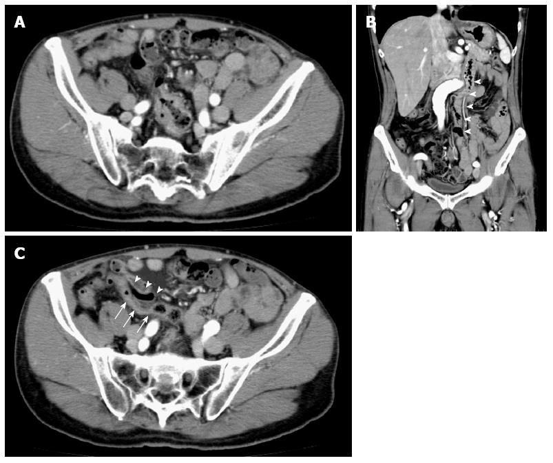
Figure 1 Computed tomography image.
A: Ascending colon cancer placed in cavity of lesser pelvis; B: Descending colon shifted to the mid-line (arrowhead); C: Sigmoid colon (arrow) placed under the ascending colon (arrowhead).
- Citation: Tsuruta A, Kawai A, Oka Y, Okumura H, Matsumoto H, Hirai T, Nakamura M. Laparoscopic right hemicolectomy for ascending colon cancer with persistent mesocolon. World J Gastroenterol 2014; 20(18): 5557-5560
- URL: https://www.wjgnet.com/1007-9327/full/v20/i18/5557.htm
- DOI: https://dx.doi.org/10.3748/wjg.v20.i18.5557


