Copyright
©2014 Baishideng Publishing Group Co.
World J Gastroenterol. May 7, 2014; 20(17): 5157-5164
Published online May 7, 2014. doi: 10.3748/wjg.v20.i17.5157
Published online May 7, 2014. doi: 10.3748/wjg.v20.i17.5157
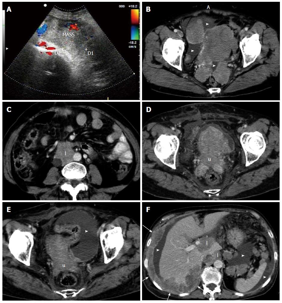
Figure 3 A 64-year-old woman with frequent urination and low back pain.
A: Abdominal US demonstrated a large heterogeneous hypoechoic mass with surrounding blood flow; B, C: Contrast-enhanced computed tomography (CT) before treatment showed multiple well-enhanced masses with variable sizes in the pelvic cavity, as well as a small amount of ascites (arrowheads) and lymphadenopathy within the retroperitoneum (l); D: Enhanced CT at the later stage of radiotherapy, showed marked shrinkage of the pelvic tumors (“u” for uterus), along with a small volume of ascites (arrowhead); E, F: Enhanced CT 3 wk after radiotherapy showed further reduced volume of the masses (“u” for uterus). At the same time, diffuse and nodular serous membrane thickening (arrows) with liver infiltration and innumerable mesenteric masses of variable size in the left upper quadrant, and lymphadenopathy (l) within the retroperitoneum and hepatic portal region, along with a moderate volume of ascites (arrowhead), were demonstrated. All of the tumor tissues presented with heterogeneous moderate enhancement.
- Citation: Shen XZ, Zhao JG, Wu JJ, Liu F. Clinical and computed tomography features of adult abdominopelvic desmoplastic small round cell tumor. World J Gastroenterol 2014; 20(17): 5157-5164
- URL: https://www.wjgnet.com/1007-9327/full/v20/i17/5157.htm
- DOI: https://dx.doi.org/10.3748/wjg.v20.i17.5157


