Copyright
©2014 Baishideng Publishing Group Co.
World J Gastroenterol. May 7, 2014; 20(17): 5157-5164
Published online May 7, 2014. doi: 10.3748/wjg.v20.i17.5157
Published online May 7, 2014. doi: 10.3748/wjg.v20.i17.5157
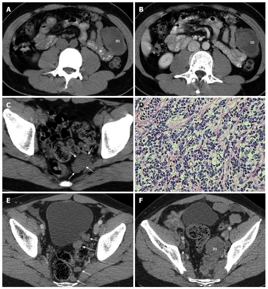
Figure 1 A 26-year-old man without any clinical symptoms.
A-C: Preoperative plain and contrast-enhanced computed tomography (CT) showed a solid-cystic mass (m) in the left mid-abdomen and a homogeneous soft-tissue mass (arrows) in the pelvis (A and C: Plain scan, B: Enhanced scan). The solid area of the abdominal tumor showed slight enhancement; D: Photomicrograph of surgical specimen showed nests or clusters of small tumor cells outlined by characteristic desmoplastic stromal bands (hematoxylin and eosin staining, × 400); E: CT re-examination 33 mo after surgery showed multiple homogeneous tumor nodules (arrows) reappearing in the retrovesical and pararectal regions; F: Six months later, CT showed that pelvic tumor nodules had enlarged rapidly forming a multinodular confluent tumor (m), as well as a slightly enlarged lymph node (arrow).
- Citation: Shen XZ, Zhao JG, Wu JJ, Liu F. Clinical and computed tomography features of adult abdominopelvic desmoplastic small round cell tumor. World J Gastroenterol 2014; 20(17): 5157-5164
- URL: https://www.wjgnet.com/1007-9327/full/v20/i17/5157.htm
- DOI: https://dx.doi.org/10.3748/wjg.v20.i17.5157


