Copyright
©2014 Baishideng Publishing Group Co.
World J Gastroenterol. May 7, 2014; 20(17): 5147-5152
Published online May 7, 2014. doi: 10.3748/wjg.v20.i17.5147
Published online May 7, 2014. doi: 10.3748/wjg.v20.i17.5147
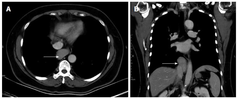
Figure 1 Computed tomography of the patient’s chest showing a well-circumscribed soft-tissue mass (white arrow), approximately 4.
35 cm × 2.5 cm × 6.14 cm in size, over the middle mediastinum, with mild compression on the esophagus. A: Sagittal view; B: Transverse view.
- Citation: Chen YY, Huang TW, Chang H, Hsu HH, Lee SC. Intrathoracic caudate lobe of the liver: A case report and literature review. World J Gastroenterol 2014; 20(17): 5147-5152
- URL: https://www.wjgnet.com/1007-9327/full/v20/i17/5147.htm
- DOI: https://dx.doi.org/10.3748/wjg.v20.i17.5147


