Copyright
©2014 Baishideng Publishing Group Co.
World J Gastroenterol. May 7, 2014; 20(17): 5141-5146
Published online May 7, 2014. doi: 10.3748/wjg.v20.i17.5141
Published online May 7, 2014. doi: 10.3748/wjg.v20.i17.5141
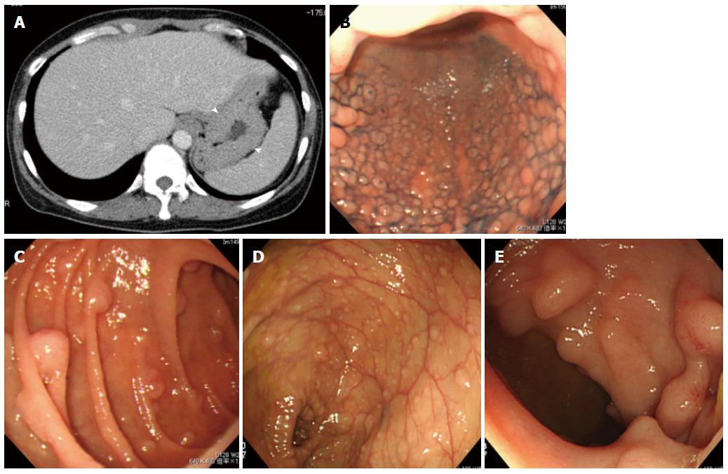
Figure 1 Images of typical type multiple lymphomatous polyposis.
A: Abdominal CT: thickening of the gastric wall (arrow head) was clearly observed; B, C: Gastrointestinal endoscopy: stomach (dye spraying) (B), descending portion of the duodenum (C); D, E: Colonoscopy: transverse colon (D), terminal ileum (E). B-E detected typical multiple lymphomatous polyposis, but none showed uptake on fluorodeoxy-glucose-positron emission tomography/computed tomography.
- Citation: Saito M, Miyazaki M, Tanino M, Tanaka S, Miyashita K, Izumiyama K, Mori A, Irie T, Tanaka M, Morioka M, Tsukamoto E. 18F-FDG PET/CT imaging for a gastrointestinal mantle cell lymphoma with multiple lymphomatous polyposis. World J Gastroenterol 2014; 20(17): 5141-5146
- URL: https://www.wjgnet.com/1007-9327/full/v20/i17/5141.htm
- DOI: https://dx.doi.org/10.3748/wjg.v20.i17.5141


