Copyright
©2014 Baishideng Publishing Group Co.
World J Gastroenterol. May 7, 2014; 20(17): 5131-5134
Published online May 7, 2014. doi: 10.3748/wjg.v20.i17.5131
Published online May 7, 2014. doi: 10.3748/wjg.v20.i17.5131
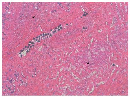
Figure 2 Pathologic examination.
Pathologic examination of the resected specimen exhibiting multiple microspheres mainly in the vessels (arrows), the microspheres were surrounded by intense fibrosis (black arrow heads) with tumor cells only on the tumor periphery (white arrow head).
- Citation: Servajean C, Gilabert M, Piana G, Monges G, Delpero JR, Brenot I, Raoul JL. One case of intrahepatic cholangiocarcinoma amenable to resection after radioembolization. World J Gastroenterol 2014; 20(17): 5131-5134
- URL: https://www.wjgnet.com/1007-9327/full/v20/i17/5131.htm
- DOI: https://dx.doi.org/10.3748/wjg.v20.i17.5131


