Copyright
©2014 Baishideng Publishing Group Co.
World J Gastroenterol. May 7, 2014; 20(17): 5066-5073
Published online May 7, 2014. doi: 10.3748/wjg.v20.i17.5066
Published online May 7, 2014. doi: 10.3748/wjg.v20.i17.5066
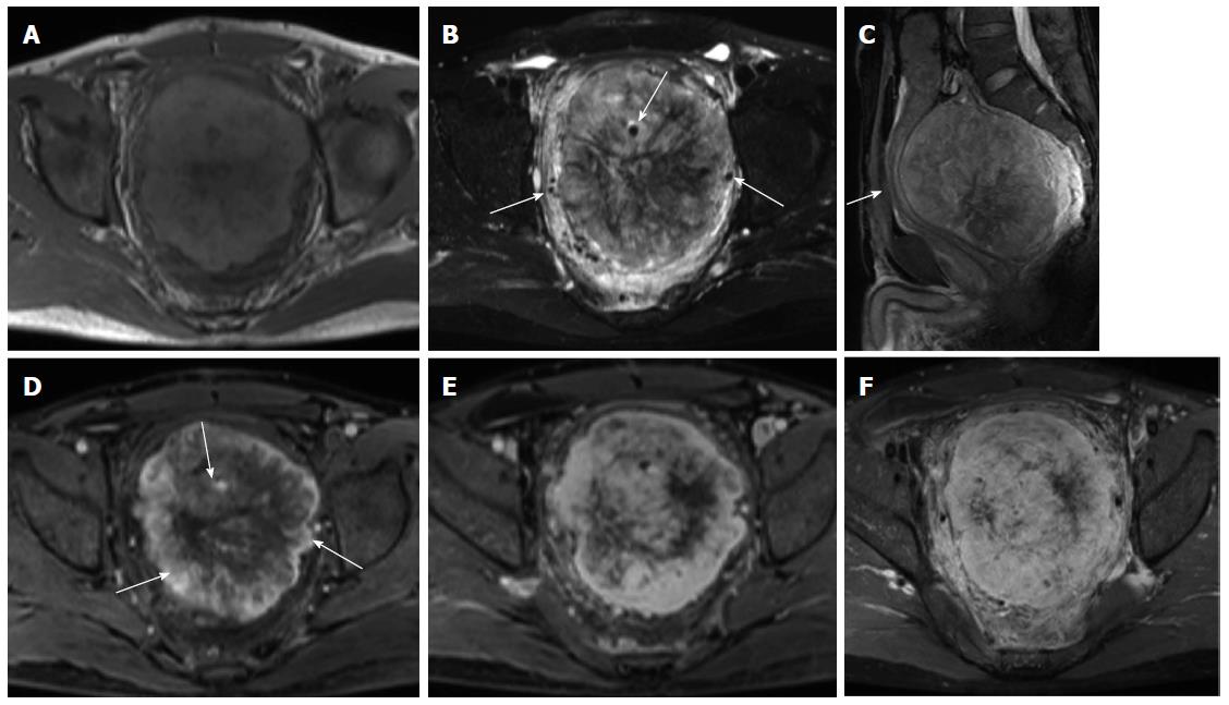
Figure 2 Magnetic resonance images of a solitary fibrous tumor in the presacral space.
A: T1-weighted Magnetic resonance (MR) image showed that the mass is lobulated and almost isointense to the muscle. Radial hypointensity is seen in the center; B: Axial fat-suppressed T2-weighted MR image shows that the mass is predominantly hyperintense with radial areas of low signal intensity. In addition, intra- and extra-tumoral flow voids can be detected (arrows); C: Sagittal fat-suppressed T2-weighted MR image showing that the rectum is compressed anteriorly by the mass (arrow); D: The mass demonstrated intense heterogeneous enhancement in the arterial phase. The feeding vessels (vascular pedicle) can be demonstrated clearly (arrows); E, F: The mass showed persistent and progressive enhancement in the portal venous (E) and delayed phase (F).
- Citation: Li XM, Reng J, Zhou P, Cao Y, Cheng ZZ, Xiao Y, Xu GH. Solitary fibrous tumors in abdomen and pelvis: Imaging characteristics and radiologic-pathologic correlation. World J Gastroenterol 2014; 20(17): 5066-5073
- URL: https://www.wjgnet.com/1007-9327/full/v20/i17/5066.htm
- DOI: https://dx.doi.org/10.3748/wjg.v20.i17.5066


