Copyright
©2014 Baishideng Publishing Group Co.
World J Gastroenterol. May 7, 2014; 20(17): 5066-5073
Published online May 7, 2014. doi: 10.3748/wjg.v20.i17.5066
Published online May 7, 2014. doi: 10.3748/wjg.v20.i17.5066
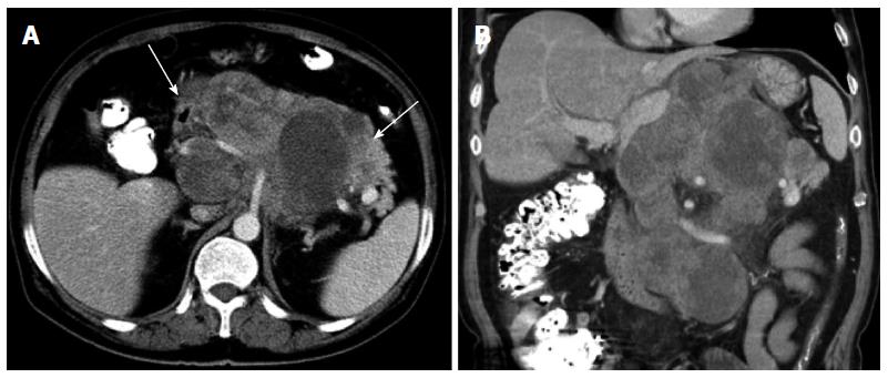
Figure 1 Computed tomography findings of a solitary fibrous tumor in the retroperitoneum.
A: Contrast-enhanced axial image showing a lobulated, ill-defined, and heterogeneously moderate enhancing mass in the retroperitoneum. The mass invades the pancreas and stomach (arrows), and the celiac trunk and its branches were enveloped; B: The contrast-enhanced coronal reconstruction image shows envelopment of the branches of the celiac trunk.
- Citation: Li XM, Reng J, Zhou P, Cao Y, Cheng ZZ, Xiao Y, Xu GH. Solitary fibrous tumors in abdomen and pelvis: Imaging characteristics and radiologic-pathologic correlation. World J Gastroenterol 2014; 20(17): 5066-5073
- URL: https://www.wjgnet.com/1007-9327/full/v20/i17/5066.htm
- DOI: https://dx.doi.org/10.3748/wjg.v20.i17.5066


