Copyright
©2014 Baishideng Publishing Group Co.
World J Gastroenterol. Apr 28, 2014; 20(16): 4811-4816
Published online Apr 28, 2014. doi: 10.3748/wjg.v20.i16.4811
Published online Apr 28, 2014. doi: 10.3748/wjg.v20.i16.4811
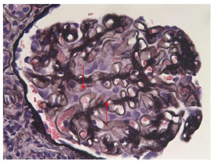
Figure 2 Optic microscopy: Jones’ staining.
Presence of spikes in a diffuse manner (arrows) (silver impregnation of the basement membrane, original magnification × 400).
- Citation: Warling O, Bovy C, Coïmbra C, Noterdaeme T, Delwaide J, Louis E. Overlap syndrome consisting of PSC-AIH with concomitant presence of a membranous glomerulonephritis and ulcerative colitis. World J Gastroenterol 2014; 20(16): 4811-4816
- URL: https://www.wjgnet.com/1007-9327/full/v20/i16/4811.htm
- DOI: https://dx.doi.org/10.3748/wjg.v20.i16.4811


