Copyright
©2014 Baishideng Publishing Group Co.
World J Gastroenterol. Apr 21, 2014; 20(15): 4462-4466
Published online Apr 21, 2014. doi: 10.3748/wjg.v20.i15.4462
Published online Apr 21, 2014. doi: 10.3748/wjg.v20.i15.4462
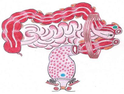
Figure 1 Details of the lesions and the procedures carried out in our patient.
The drawing shows the staple line anastomosis of the STARR with the peristaples fibrosis (grey) triggering the nerve spindles (black dots) located above the puborectalis muscle and the levator ani. The blue circles indicate the deep rectal and colonic biopsies, aimed at evaluating the intrinsic nerves. The red spots in the rectum indicate the diversion proctitis and the asterix on the small bowel loop indicates the parastomal hernia, due to the diastasis of the abdominal wall muscle.
- Citation: Pescatori LC, Villanacci V, Pescatori M. Failed stapled rectal resection in a constipated patient with rectal aganglionosis. World J Gastroenterol 2014; 20(15): 4462-4466
- URL: https://www.wjgnet.com/1007-9327/full/v20/i15/4462.htm
- DOI: https://dx.doi.org/10.3748/wjg.v20.i15.4462


