Copyright
©2014 Baishideng Publishing Group Co.
World J Gastroenterol. Apr 21, 2014; 20(15): 4457-4461
Published online Apr 21, 2014. doi: 10.3748/wjg.v20.i15.4457
Published online Apr 21, 2014. doi: 10.3748/wjg.v20.i15.4457
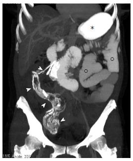
Figure 3 Multi-detector contrast-enhanced computed tomography.
An 80 mm thick maximum intensity projection (MIP) reconstructed image is shown; The laparotomy sponge (arrow-heads) is clearly depicted in the right flank extending from the ileo-cecal valve to the proximal transverse colon where the perforation occurred; Stomach (*) and jejunal loops (°) appear opacified by orally administered iodinated contrast medium (Gastrografin).
- Citation: Camera L, Sagnelli M, Guadagno P, Mainenti PP, Marra T, Scotto di Santolo M, Fei L, Salvatore M. Colonic perforation by a transmural and transvalvular migrated retained sponge: Multi-detector computed tomography findings. World J Gastroenterol 2014; 20(15): 4457-4461
- URL: https://www.wjgnet.com/1007-9327/full/v20/i15/4457.htm
- DOI: https://dx.doi.org/10.3748/wjg.v20.i15.4457


