Copyright
©2014 Baishideng Publishing Group Co.
World J Gastroenterol. Apr 21, 2014; 20(15): 4457-4461
Published online Apr 21, 2014. doi: 10.3748/wjg.v20.i15.4457
Published online Apr 21, 2014. doi: 10.3748/wjg.v20.i15.4457
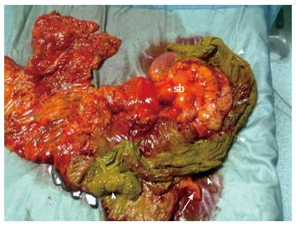
Figure 2 Right hemi-colectomy.
A huge laparotomy sponge (arrow-heads) can be seen on dissection of the surgical specimen which included a cluster of small bowel loops (sb) and the appendix (arrow).
- Citation: Camera L, Sagnelli M, Guadagno P, Mainenti PP, Marra T, Scotto di Santolo M, Fei L, Salvatore M. Colonic perforation by a transmural and transvalvular migrated retained sponge: Multi-detector computed tomography findings. World J Gastroenterol 2014; 20(15): 4457-4461
- URL: https://www.wjgnet.com/1007-9327/full/v20/i15/4457.htm
- DOI: https://dx.doi.org/10.3748/wjg.v20.i15.4457


