Copyright
©2014 Baishideng Publishing Group Co.
World J Gastroenterol. Apr 21, 2014; 20(15): 4453-4456
Published online Apr 21, 2014. doi: 10.3748/wjg.v20.i15.4453
Published online Apr 21, 2014. doi: 10.3748/wjg.v20.i15.4453
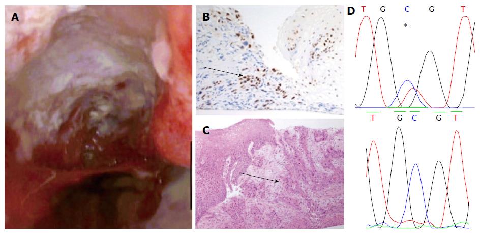
Figure 2 Endoscopic, genetic and histological figures of the patient after radiofrequency ablation.
A: Endoscopic view of a 4cm ulcerating mass between 35 to 39 cm; B: Example of positive p63 stain of the biopsies taken from the ulcerating mass (arrow); C: Haematoxylin and eosin stain of biopsies taken from the ulcer crater demonstrating invasive squamous cell cancer (arrow); D: Sanger sequencing of a mutation in TP53 c.817 C>T p. R273C (upper panel, starred) Wild type Sanger sequence of TP53 (lower panel).
- Citation: Zeki SS, Haidry R, Justo-Rodriguez M, Lovat LB, Wright NA, McDonald SA. Squamous cell carcinoma after radiofrequency ablation for Barrett's dysplasia. World J Gastroenterol 2014; 20(15): 4453-4456
- URL: https://www.wjgnet.com/1007-9327/full/v20/i15/4453.htm
- DOI: https://dx.doi.org/10.3748/wjg.v20.i15.4453


