Copyright
©2014 Baishideng Publishing Group Co.
World J Gastroenterol. Apr 21, 2014; 20(15): 4433-4439
Published online Apr 21, 2014. doi: 10.3748/wjg.v20.i15.4433
Published online Apr 21, 2014. doi: 10.3748/wjg.v20.i15.4433
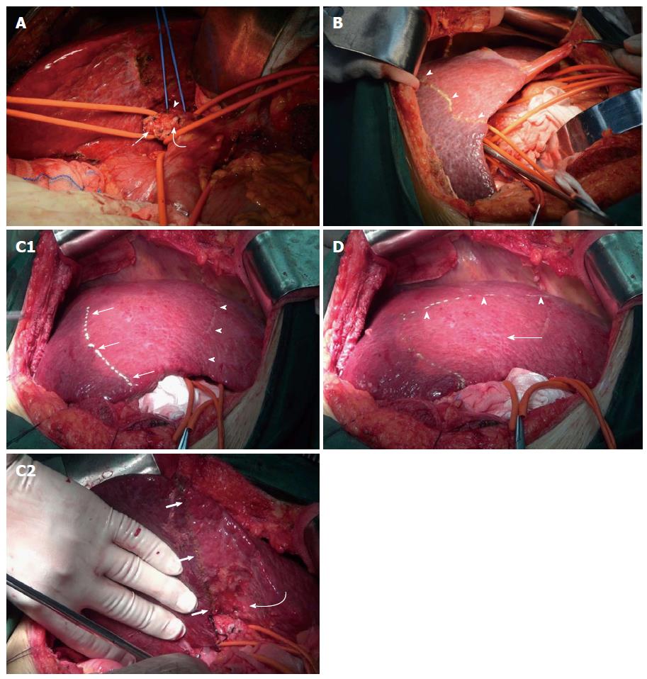
Figure 2 Surgical procedures.
A: Right hemihepatic Glissonean pedicle and segment 6 and 7 Glissonean pedicle in Case 4 were sequentially divided. Arrow: Segment 6 and 7 Glissonean pedicle; arrowhead: Segment 5 and 8 Glissonean pedicle; curved arrow: Right hemihepatic Glissonean pedicle; B: After occlusion of the right hemihepatic Glissonean pedicle in Case 4, the right liver showed obvious ischemia. The demarcation between the right and left liver could be easily determined (arrowheads); C: After the segment 6 and 7 Glissonean pedicle of Case 4 was ligated, segments 6 and 7 showed obvious ischemia. The interface between segments 6 and 5 could be easily demarcated. Arrow: Demarcation between segments 6 and 5 upon the diaphragmatic surface of the liver; Arrowheads: Demarcation between the right and left liver; Bold arrows: Demarcation between segments 6 and 5 on the visceral surface of the liver; Curved arrow: Fossa of gallbladder (C1, C2); D: Segment 5 was determined and a “┏┛” shaped broken resection line was marked upon the diaphragmatic surface of the liver in Case 4. Arrowheads: Demarcation between segments 5 and 8; Arrow: Segment 5.
- Citation: Jia CK, Weng J, Chen YK, Fu Y. Anatomic resection of liver segments 6-8 for hepatocellular carcinoma. World J Gastroenterol 2014; 20(15): 4433-4439
- URL: https://www.wjgnet.com/1007-9327/full/v20/i15/4433.htm
- DOI: https://dx.doi.org/10.3748/wjg.v20.i15.4433


