Copyright
©2014 Baishideng Publishing Group Co.
World J Gastroenterol. Apr 21, 2014; 20(15): 4433-4439
Published online Apr 21, 2014. doi: 10.3748/wjg.v20.i15.4433
Published online Apr 21, 2014. doi: 10.3748/wjg.v20.i15.4433
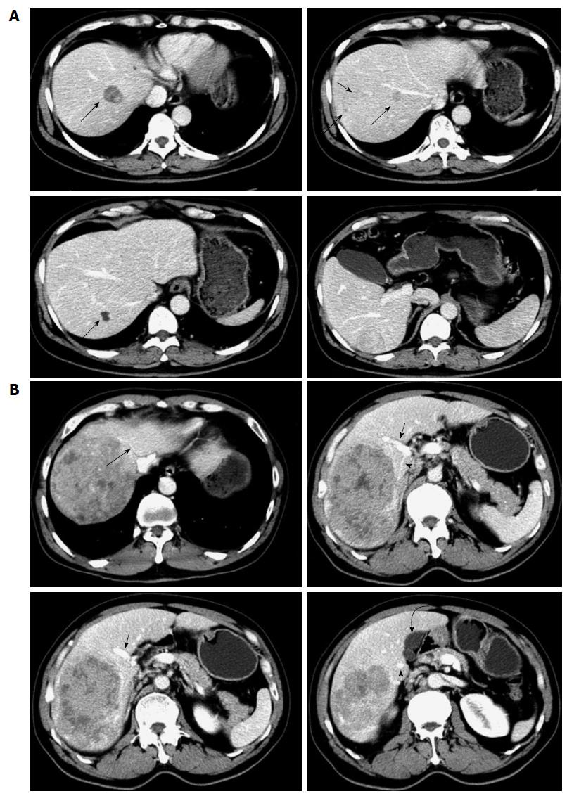
Figure 1 Preoperative imaging showed that segment 5 was free of tumor in all patients.
A: Multiple tumors were found in segments 6, 7 and 8 by contrast-enhanced computed tomography scans (arrows) in Case 1; B: Huge tumor located in the right liver in Case 4. Arrow: Middle hepatic vein; Arrowhead: Right anterior portal vein; Triangular arrow: Right posterior portal vein; Curved arrow: Gallbladder.
- Citation: Jia CK, Weng J, Chen YK, Fu Y. Anatomic resection of liver segments 6-8 for hepatocellular carcinoma. World J Gastroenterol 2014; 20(15): 4433-4439
- URL: https://www.wjgnet.com/1007-9327/full/v20/i15/4433.htm
- DOI: https://dx.doi.org/10.3748/wjg.v20.i15.4433


