Copyright
©2014 Baishideng Publishing Group Co.
World J Gastroenterol. Apr 21, 2014; 20(15): 4244-4255
Published online Apr 21, 2014. doi: 10.3748/wjg.v20.i15.4244
Published online Apr 21, 2014. doi: 10.3748/wjg.v20.i15.4244
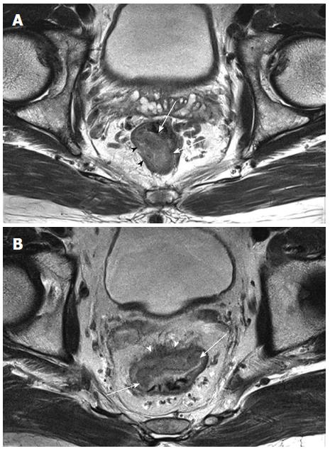
Figure 2 Stage T2 and T3 rectal cancer detected by magnetic resonance imaging.
A: T2-weighted magnetic resonance image shows an intraluminal polypoid mass (arrows) confined to the intact, hypo-intense muscularis propria (arrowheads), suggestive of a T2 cancer; B: T3 lesion is seen as a tumor (arrows) that extends through the hypo-intense muscle layer into the perirectal fat (arrowheads).
- Citation: Heo SH, Kim JW, Shin SS, Jeong YY, Kang HK. Multimodal imaging evaluation in staging of rectal cancer. World J Gastroenterol 2014; 20(15): 4244-4255
- URL: https://www.wjgnet.com/1007-9327/full/v20/i15/4244.htm
- DOI: https://dx.doi.org/10.3748/wjg.v20.i15.4244


