Copyright
©2014 Baishideng Publishing Group Co.
World J Gastroenterol. Apr 14, 2014; 20(14): 4001-4010
Published online Apr 14, 2014. doi: 10.3748/wjg.v20.i14.4001
Published online Apr 14, 2014. doi: 10.3748/wjg.v20.i14.4001
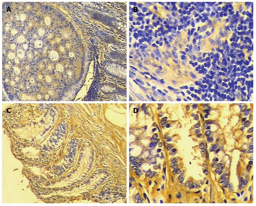
Figure 1 Immunohistochemistry of epithelial membrane protein 1 protein in colorectal carcinoma and adjacent normal tissue.
A and B: Representative sample of colorectal carcinoma (A: SP × 100, B: SP × 400). There is little staining for epithelial membrane protein 1 (EMP1); C and D: normal colorectal tissue (C: SP × 100, D: SP × 400). There is intense yellow and yellow-brown staining of EMP1.
- Citation: Sun GG, Wang YD, Cui DW, Cheng YJ, Hu WN. Epithelial membrane protein 1 negatively regulates cell growth and metastasis in colorectal carcinoma. World J Gastroenterol 2014; 20(14): 4001-4010
- URL: https://www.wjgnet.com/1007-9327/full/v20/i14/4001.htm
- DOI: https://dx.doi.org/10.3748/wjg.v20.i14.4001


