Copyright
©2014 Baishideng Publishing Group Co.
World J Gastroenterol. Apr 14, 2014; 20(14): 3795-3803
Published online Apr 14, 2014. doi: 10.3748/wjg.v20.i14.3795
Published online Apr 14, 2014. doi: 10.3748/wjg.v20.i14.3795
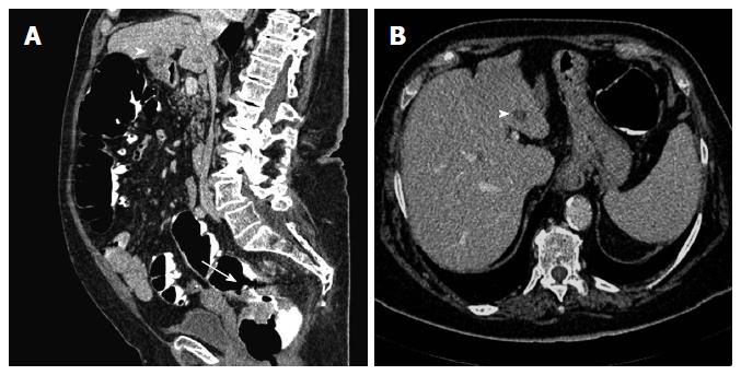
Figure 6 Computed tomography colonography of T3 M1 rectal cancer.
A: A sagittal reconstructed image shows the rectal tumor (arrow) and a hepatic metastasis appearing as a hypoattenuating focal lesion (arrowhead); B: An axial source image demonstrates that the metastatic lesion is located in the third hepatic segment (arrowhead).
- Citation: Sali L, Falchini M, Taddei A, Mascalchi M. Role of preoperative CT colonography in patients with colorectal cancer. World J Gastroenterol 2014; 20(14): 3795-3803
- URL: https://www.wjgnet.com/1007-9327/full/v20/i14/3795.htm
- DOI: https://dx.doi.org/10.3748/wjg.v20.i14.3795


