Copyright
©2014 Baishideng Publishing Group Co.
World J Gastroenterol. Apr 14, 2014; 20(14): 3795-3803
Published online Apr 14, 2014. doi: 10.3748/wjg.v20.i14.3795
Published online Apr 14, 2014. doi: 10.3748/wjg.v20.i14.3795
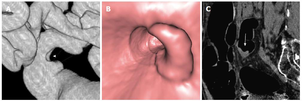
Figure 3 Computed tomography colonography of a T2 vegetating lesion of the sigmoid colon.
A: A Computed tomography colonography (CTC) SSD reconstructed image shows a trapezoid type wall deformity of the sigmoid colon (arrowhead); B: A CTC endoluminal image demonstrates that wall involvement is less than 50%of luminal circumference. C: A CTC sagittal reconstructed image shows that the lesion (arrow) has sharp margins.
- Citation: Sali L, Falchini M, Taddei A, Mascalchi M. Role of preoperative CT colonography in patients with colorectal cancer. World J Gastroenterol 2014; 20(14): 3795-3803
- URL: https://www.wjgnet.com/1007-9327/full/v20/i14/3795.htm
- DOI: https://dx.doi.org/10.3748/wjg.v20.i14.3795


