Copyright
©2014 Baishideng Publishing Group Co.
World J Gastroenterol. Apr 14, 2014; 20(14): 3795-3803
Published online Apr 14, 2014. doi: 10.3748/wjg.v20.i14.3795
Published online Apr 14, 2014. doi: 10.3748/wjg.v20.i14.3795
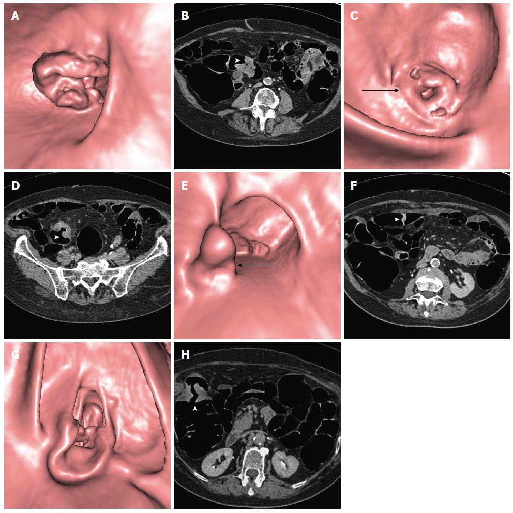
Figure 1 Computed tomography colonography performed in an 83-year-old female with incomplete colonoscopy due to stenosing adenocarcinoma of the sigmoid colon.
Computed tomography colonography (CTC) identified three other synchronous colonic tumors confirmed as adenocarcinomas at surgery. CTC endoluminal and axial source images are shown. A, B: Distal stenosing lesion of the sigmoid (arrowhead); C, D: Proximal stenosing lesion of the sigmoid (arrow and arrowhead); E, F: Vegetating lesion of the transverse colon (arrow and arrowhead); G, H: Stenosing lesion of the right flexure (arrowhead).
- Citation: Sali L, Falchini M, Taddei A, Mascalchi M. Role of preoperative CT colonography in patients with colorectal cancer. World J Gastroenterol 2014; 20(14): 3795-3803
- URL: https://www.wjgnet.com/1007-9327/full/v20/i14/3795.htm
- DOI: https://dx.doi.org/10.3748/wjg.v20.i14.3795


