Copyright
©2014 Baishideng Publishing Group Co.
World J Gastroenterol. Apr 14, 2014; 20(14): 3719-3737
Published online Apr 14, 2014. doi: 10.3748/wjg.v20.i14.3719
Published online Apr 14, 2014. doi: 10.3748/wjg.v20.i14.3719
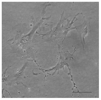
Figure 6 Hepatic stellate cells with their cytoplasmic processes, after 4 d in culture (authors’ archive).
Light microscopy (magnification × 200). Scale bar = 100 μm.
- Citation: Paschos KA, Majeed AW, Bird NC. Natural history of hepatic metastases from colorectal cancer - pathobiological pathways with clinical significance. World J Gastroenterol 2014; 20(14): 3719-3737
- URL: https://www.wjgnet.com/1007-9327/full/v20/i14/3719.htm
- DOI: https://dx.doi.org/10.3748/wjg.v20.i14.3719


