Copyright
©2014 Baishideng Publishing Group Co.
World J Gastroenterol. Apr 7, 2014; 20(13): 3597-3608
Published online Apr 7, 2014. doi: 10.3748/wjg.v20.i13.3597
Published online Apr 7, 2014. doi: 10.3748/wjg.v20.i13.3597
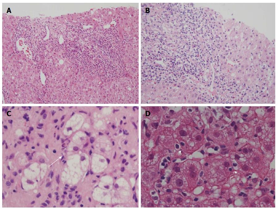
Figure 1 Histological features of primary biliary cirrhosis with interface hepatitis and autoimmune hepatitis.
A: Moderate interface hepatitis (score 3) in autoimmune hepatitis (AIH); B: Moderate interface hepatitis (score 3) in primary biliary cirrhosis (PBC); C: Rosette formation; regenerative hepatocytes arranged around a bile canaliculus (arrow), found in AIH; D: Emperipolesis, the engulfment of lymphocytes within hepatocytes (arrowheads), was found in areas of interface hepatitis. Hematoxylin and eosin stain, original magnifications × 200 ( A, B); × 600 (C, D).
- Citation: Kobayashi M, Kakuda Y, Harada K, Sato Y, Sasaki M, Ikeda H, Terada M, Mukai M, Kaneko S, Nakanuma Y. Clinicopathological study of primary biliary cirrhosis with interface hepatitis compared to autoimmune hepatitis. World J Gastroenterol 2014; 20(13): 3597-3608
- URL: https://www.wjgnet.com/1007-9327/full/v20/i13/3597.htm
- DOI: https://dx.doi.org/10.3748/wjg.v20.i13.3597


