Copyright
©2014 Baishideng Publishing Group Co.
World J Gastroenterol. Apr 7, 2014; 20(13): 3590-3596
Published online Apr 7, 2014. doi: 10.3748/wjg.v20.i13.3590
Published online Apr 7, 2014. doi: 10.3748/wjg.v20.i13.3590
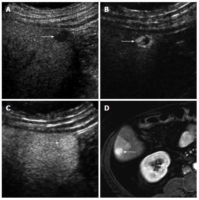
Figure 6 A 53-year-old woman with hemangioma.
A: Gray-scale ultrasound shows a hypoechoic nodule (arrow) in the subcapsular portion of the liver; B: There is a peripheral strong hyperenhancement (arrows) in the arterial phase of contrast-enhanced ultrasound; C: The nodule (arrow) is homogeneously hyperechoic to the liver in the portal venous phase; D: The nodule (arrow) is homogeneously hyperintense to the liver without the appearance of peripheral enhancement in the arterial phase of contrast-enhanced T1-weighed magnetic resonance image.
- Citation: Kim TK, Jang HJ. Contrast-enhanced ultrasound in the diagnosis of nodules in liver cirrhosis. World J Gastroenterol 2014; 20(13): 3590-3596
- URL: https://www.wjgnet.com/1007-9327/full/v20/i13/3590.htm
- DOI: https://dx.doi.org/10.3748/wjg.v20.i13.3590


