Copyright
©2014 Baishideng Publishing Group Co.
World J Gastroenterol. Apr 7, 2014; 20(13): 3590-3596
Published online Apr 7, 2014. doi: 10.3748/wjg.v20.i13.3590
Published online Apr 7, 2014. doi: 10.3748/wjg.v20.i13.3590
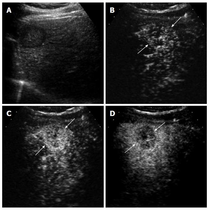
Figure 3 A 70-year-old man with intrahepatic cholangiocarcinoma detected during surveillance for hepatocellular carcinoma.
A: Gray-scale ultrasound shows a hypoechoic mass in the liver; B: Contrast-enhanced ultrasound (CEUS) in the arterial phase obtained 14 s after contrast injection shows mild hypervascularity in the periphery of the mass (arrows); C: CEUS obtained at 19 s after contrast injection shows diffuse hypervascularity of the mass (arrows); D: The mass (arrows) shows rapid washout at 28 s after contrast injection. Biopsy revealed cholangiocarcinoma.
- Citation: Kim TK, Jang HJ. Contrast-enhanced ultrasound in the diagnosis of nodules in liver cirrhosis. World J Gastroenterol 2014; 20(13): 3590-3596
- URL: https://www.wjgnet.com/1007-9327/full/v20/i13/3590.htm
- DOI: https://dx.doi.org/10.3748/wjg.v20.i13.3590


