Copyright
©2014 Baishideng Publishing Group Co.
World J Gastroenterol. Apr 7, 2014; 20(13): 3590-3596
Published online Apr 7, 2014. doi: 10.3748/wjg.v20.i13.3590
Published online Apr 7, 2014. doi: 10.3748/wjg.v20.i13.3590
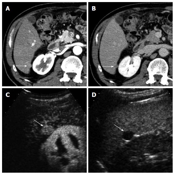
Figure 2 A 63-year-old man with moderately-differentiated hepatocellular carcinoma.
A: Computed tomography scan in the arterial phase shows a small exophytic nodule (arrow) which shows similar attenuation to the liver; B: The nodule (arrow) is slightly hypoattenuating to the liver in the delayed phase; C: Contrast-enhanced ultrasound (CEUS) in the arterial phase clearly demonstrates hypervascularity of the nodule (arrow); D: CEUS in the portal venous phase shows washout of the nodule (arrow).
- Citation: Kim TK, Jang HJ. Contrast-enhanced ultrasound in the diagnosis of nodules in liver cirrhosis. World J Gastroenterol 2014; 20(13): 3590-3596
- URL: https://www.wjgnet.com/1007-9327/full/v20/i13/3590.htm
- DOI: https://dx.doi.org/10.3748/wjg.v20.i13.3590


