Copyright
©2014 Baishideng Publishing Group Co.
World J Gastroenterol. Apr 7, 2014; 20(13): 3590-3596
Published online Apr 7, 2014. doi: 10.3748/wjg.v20.i13.3590
Published online Apr 7, 2014. doi: 10.3748/wjg.v20.i13.3590
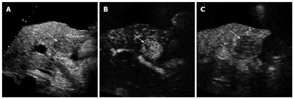
Figure 1 A 61-year-old man with hepatocellular carcinoma and renal failure.
A: Gray-scale ultrasound shows a hypoechoic mass (arrow) within the cirrhotic liver. There is a large amount of ascites surrounding the liver. Contrast-enhanced computed tomography or magnetic resonance scan could not be performed because of renal failure; B: Contrast-enhanced ultrasound (CEUS) image in the arterial phase shows hypervascularity of the mass (arrow) relative to the liver; C: The mass (arrow) shows washout in the portal venous phase. The diagnosis of hepatocellular carcinoma was made based on imaging findings of CEUS without biopsy.
- Citation: Kim TK, Jang HJ. Contrast-enhanced ultrasound in the diagnosis of nodules in liver cirrhosis. World J Gastroenterol 2014; 20(13): 3590-3596
- URL: https://www.wjgnet.com/1007-9327/full/v20/i13/3590.htm
- DOI: https://dx.doi.org/10.3748/wjg.v20.i13.3590


